Topic:
Fluorescence microscopy is a widely used tool for monitoring cellular physiology. The absorption and subsequent re-radiation of light by organic and inorganic specimens is typically the result of well-established physical phenomena described as fluorescence.
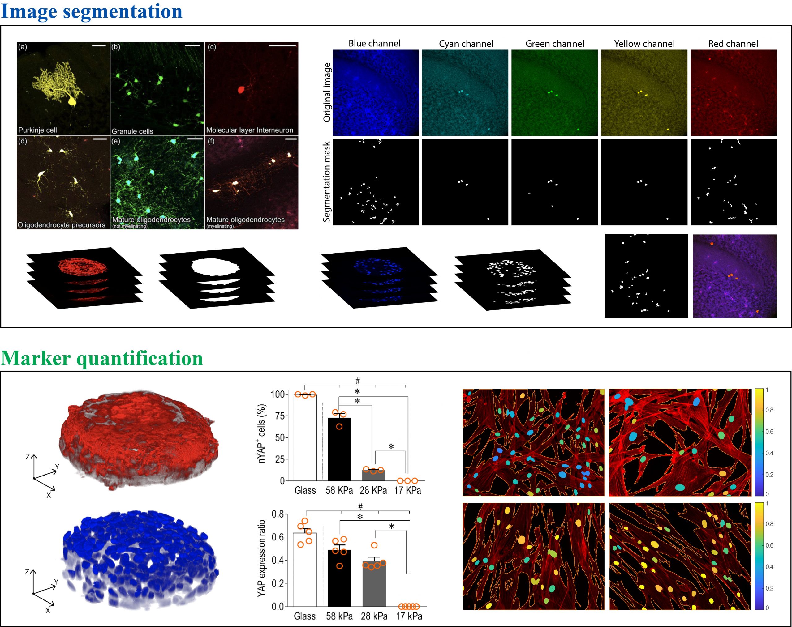
The technological advancements in multicolor labelling of cells and tissues, in association with modern semi-automated or automated microscopy, produce enormous sets of data. Looking at the resulting images by eye is extremely time consuming and subjectivity may introduce biases and errors in the analysis. Thus, many biologists find they need software to analyze images easily, accurately and objectively. This line of research is focused on the development of automated algorithms for the detection and segmentation of cellular structures, resulting in the quantification of fluorescent markers.