Year projects were funded:

2020 - 2024

Effects of bilateral subthalamic nucleus deep brain stimulation on motor control strategies of patients affected by advanced Parkinson's disease during dual-task walking (PD-DBS)

Description:

Deep Brain Stimulation of the SubThalamic Nucleus(STN-DBS) is a well-established surgical therapy for patients with advanced Parkinson’s disease (PD) and motor complications that cannot be adequately managed with medication. Instrumented gait analysis already proved successful for objectively evaluating alterations of locomotor patterns in a wide variety of neurological and neurodegenerative diseases, including PD. Only recently, the study of motor modules, through the muscle synergy theory, revealed its potential in clinics for understanding the basic mechanisms through which the Central Nervous System (CNS) coordinates different motor tasks. In particular, it is possible to reconstruct the group of muscles that synergistically cooperate and the time-dependent neural command that drive it. The aims of this project are: (1) evaluating the effect of bilateral STN-DBS on the motor control strategies of PD patients during dual-task walking, (2) defining and validating a clinical protocol to quantify the functional changes of PD patients through a complete gait and muscle synergy analysis. The assessment will be conducted before STN-DBS surgery, and at 3 months and at 12 months after it, by recording non-invasively the electromyographic (sEMG) activity of the main muscles involved in gait. New medical knowledge will be acquired for the management of PD patients.

Scientific Director:
  • Valentina Agostini (Associate Professor)
  • Year: 2023-2025
    Other companies/universities involved in the project:
  • Prof. Michele Rosario Maria Lanotte, Dr. Laura Rizzi - Department of Neuroscience “Rita Levi Montalcini”, University of Turin, Turin
  • ERC panels:
  • ERC_LS5_11 Neurological and neurodegenerative disorders
  • PE6_11 Machine learning, statistical data processing and applications using signal processing (e.g., speech, image, video)
  • Sustainable Development Goals (SDG):
  • SDG 3 - “Ensure healthy lives and promote well-being for all at all ages”
  • People involved:
  • Valentina Agostini (Associate Professor)
  • Marco Ghislieri (Assistant Professor with time contract (RTD/a))

  • AI-VASCUES : quAntitative Imaging of VAScular dysregulation as a funCtional basis for aUtoimmune disordErs and tumorS

    Description:

    A well-known biomarker of both oncological, autoimmune and inflammatory
    diseases is their vascular “fingerprint”. The regulation of vascular function in response to changing metabolic needs is essential for the maintenance of normal tissue and organ functions. In fact, one hallmark of cancer is the deregulation of angiogenic signaling, resulting in a process called neoangiogenesis, where the newly developed blood vessels are often organized chaotically and densely-packed around the tumor. Similarly, vascular dysregulation is also involved when considering autoimmune diseases and inflammation, which is an important pathophysiological
    event that involves changes in cell composition and molecular
    characteristics, by promoting the infiltration of inflammatory immune
    cells and local oxidative stress and by triggering vascular pathological signals. Photoacoustics (PA) is a rapidly growing imaging modality that allows a non-invasive quantitative and metabolic analysis of vasculature. Studies have shown great potentials for this imaging modality, which is however still limited to ad-hoc specialized geometrical detectors or artefact-ridden images when using traditional linear probes for detection.
    The main goal of this project concerns with quantitative imaging of vasculature, and in particular vascular dysregulation, as a functional basis for autoimmune diseases and tumors using photoacoustic imaging and artificial intelligence. Innovative methods for PA volume imaging using a linear ultrasound probe will be developed and innovative multi-angle illumination of the imaged area will be explored for high-resolution and artefact-reduced volumetric and functional PA imaging. The project begins at in-silico and in-vitro levels, including simulations and phantom studies. Then, the activity will advance towards in-vivo chicken embryo studies. The project climaxes at the in-vivo clinical study of cancerous dermatological lesions and inflammation of the Achilles tendon. Importantly, artificial intelligence-based methods, specifically generative adversarial networks (GANs), will be employed to optimize and enhance the acquired photoacoustic images. Artificial intelligence methods will be employed to automatically segment and classify the acquired photoacoustic volumes and the dermoscopic and histopathological biopsy images acquired in routine clinical practice. As a result of this project, an innovative 3D photoacoustic imaging system using a common ultrasound linear probe for
    detection will be developed, making the entire system potentially miniaturizable in the future for point-of-care solutions. The partnership of AI-VASCUES is a multidisciplinary team of experts in biomedical engineering, signal and image processing, together with clinicians in two fields. This composition will provide an ideal synergy of background knowledge, tech know-how, case scenarios and outreach potential for the best potential scientific and socioeconomic impact.

    Scientific Director:
  • Kristen Meiburger (Tenure-track Assistant Professor (RTD/b))
  • Year: 2023
    Other companies/universities involved in the project:
  • Politecnico di Bari - Prof. Vitoantonio Bevilacqua
  • Prof. Marco Minetto - Università degli Studi di Torino
  • Università degli Studi di Bari Aldo Moro - Prof. Paolo Romita
  • ERC panels:
  • PE7_11 Components and systems for applications
  • ERC PE8_13 - Industrial Bioengineering
  • ERC LS7_14 - Digital medicine, e-medicine, medical applications of artificial intelligence
  • PE6_11 - Machine learning, statistical data processing and applications using signal processing (e.g. speech, image, video)
  • Sustainable Development Goals (SDG):
  • SDG 3 - “Ensure healthy lives and promote well-being for all at all ages”
  • People involved:
  • Filippo Molinari (Full Professor)
  • Silvia Seoni (Research Assistant)
  • Kristen Meiburger (Tenure-track Assistant Professor (RTD/b))
  • Bruna Cotrufo (Ph.D. student)

  • DIPAT3-Care: Digital Pathology Pathways to improve Patient Care

    Description:

    DIPAT³-Care is a funded project that aims to revolutionize the diagnosis and classification of lymphomas through artificial intelligence. The project specifically focuses on Diffuse Large B-Cell Lymphoma (DLBCL), the most common type of non-Hodgkin lymphoma, which despite being treated as a single disease, represents a heterogeneous group of tumors with varying clinical outcomes and responses to therapy.
    The core innovation of DIPAT³-Care lies in its unique approach to combining artificial intelligence with digital pathology. The project develops advanced AI tools that can analyze digitized tissue slides and integrate this analysis with molecular and clinical data. This integration aims to create a more precise and personalized approach to lymphoma diagnosis and prognosis prediction.
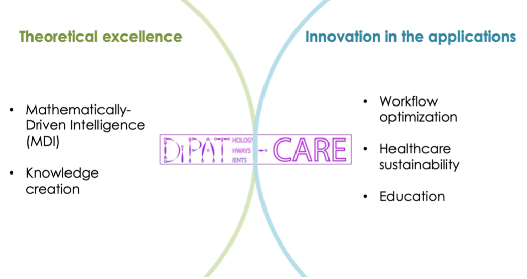
    A key technological breakthrough of the project is the development of Mathematically-Driven Intelligence (MDI), a new framework that combines traditional mathematical and statistical methods with modern AI approaches. This hybrid approach ensures higher accuracy and adaptability compared to conventional deep learning methods alone. The project also introduces solutions in stain normalization and automated tissue analysis, making the technology more reliable and reproducible across different laboratories.
    A distinctive feature of the project is its focus on explainable AI, ensuring that the automated analysis provides clear reasoning for its decisions – a crucial aspect for clinical adoption. The system will include visual aids such as heat maps and similarity comparisons to help pathologists understand and verify the AI’s conclusions.
    The project benefits from one of the largest cohorts of DLBCL cases collected worldwide, through collaboration with prestigious institutions including the European Institute of Oncology (IEO) and the University Hospital “Città della Salute e della Scienza” of Turin.

    Scientific Director:
  • Filippo Molinari (Full Professor)
  • Year: 2023
    ERC panels:
  • ERC LS7_14 - Digital medicine, e-medicine, medical applications of artificial intelligence
  • Sustainable Development Goals (SDG):
  • SDG 3 - “Ensure healthy lives and promote well-being for all at all ages”
  • People involved:
  • Massimo Salvi (Assistant Professor with time contract (RTD/a))
  • Nicola Michielli (Research Assistant)
  • Alen Shahini (Ph.D. student)

  • ImPACT-AI: Development of inclusive quantitative photoacoustic imaging solutions enabled by ethical artificial intelligence

    Description:

    Photoacoustics (PA) is a rapidly emerging imaging modality thanks to its inherent functional and metabolic capacity, sensitivity, depth penetration, non-invasiveness and radiation-free measurement of optical tissue properties. Hemoglobin is an active absorber of light and hence an ideal chromophore that enables quantitative PA-based analysis of vasculature in terms of morphology, network complexity and functional metabolism, i.e., blood oxygenation. Current limitations to PA imaging include artefact-ridden images when using traditional linear ultrasound probes (LUPs) for detection, or the need of ad-hoc detectors. Another challenge for PA
    imaging is that variations in skin tone, hence melanin content, can alter measurements between subjects in ways that the imaging device was not designed to anticipate. If ignored, this produces inaccurate functional information such as blood oxygenation levels based simply on the different skin color.

    ImPACT-AI aims to provide new PA imaging tools and artificial intelligence (AI) digital solutions for the inclusive quantitative imaging of vasculature in terms of complexity and functional oxygenation saturation analysis. Innovative methods for PA volume imaging using a LUP will be developed and innovative multi-angle illumination of the imaged area will be explored for high-resolution and artefact-reduced volumetric and functional PA imaging.
    The artificial intelligence methods will be developed by strictly following an “ethics by design” approach. AI methods, such as generative adversarial networks (GANs) and fully convolutional networks (FCNs), will be employed to optimize and enhance the acquired photoacoustic images. AI methods will be employed to automatically provide morphological and metabolic information about the acquired photoacoustic volumes. As a result of this project, new knowledge will be gained on how AI can enhance and
    enable photoacoustic imaging systems using a common LUP for detection and how an ethical and inclusive development process of AI techniques can compensate potential biases that could emerge due to skin tone variations.
    The project begins at an in-silico level designing, developing, and testing the new methodologies on simulated data. Then, the activity will advance towards in-vitro studies that consider phantoms. We will develop customized phantoms in compliance with the criteria defined by the International Photoacoustic Standardisation Consortium (IPASC). Our aim is the development of a reliable and low-cost platform that may even outclass the use of animals in terms of costs, repeatability, reproducibility, and serve as ground-truth to the AI solutions.

    Scientific Director:
  • Kristen Meiburger (Tenure-track Assistant Professor (RTD/b))
  • Year: 2023
    Other companies/universities involved in the project:
  • Politecnico di Bari - Prof. Marilena Giglio
  • Istituto di Fisica Applicata “Nello Carrara” - CNR - Lucia Cavigli
  • ERC panels:
  • PE7_11 Components and systems for applications
  • ERC PE6_7 : Artificial intelligence, intelligent systems, natural language processing
  • PE6_11 - Machine learning, statistical data processing and applications using signal processing (e.g. speech, image, video)
  • ERC LS7_14 - Digital medicine, e-medicine, medical applications of artificial intelligence
  • Sustainable Development Goals (SDG):
  • SDG 3 - “Ensure healthy lives and promote well-being for all at all ages”
  • People involved:
  • Filippo Molinari (Full Professor)
  • Silvia Seoni (Research Assistant)
  • Kristen Meiburger (Tenure-track Assistant Professor (RTD/b))
  • Bruna Cotrufo (Ph.D. student)

  • Objective monitoring of axial symptoms in Parkinson's disease: quantitative assessment in daily life based on the use of wearables, video sensing and artificial intelligence (OMNIA-PARK)

    Description:

    Parkinson’s disease (PD) is the second most prevalent neurodegenerative disorder in the aging population. It is characterized by many motor and non-motor symptoms, with a different impact on patients’ daily life activities, independence, and quality of life. In addition to the typical cardinal motor symptoms, namely bradykinesia, rigidity, and tremor, more commonly affecting the limbs, another group of motor symptoms involving the axis of the body and called axial motor symptoms comes out during the course of the disease. These symptoms proved to be main determinants of reduced independence and quality of life, and can also be a cause of mortality as the primary source of falls and aspiration pneumonia. Moreover, axial symptoms are poorly or not responsive to common therapeutic strategies and are often recognized when their consequences appear, like falls or aspiration pneumonia. A big effort of the research community in the last years has pointed toward the development of e-Health tools for a more granular and accurate detection and quantification of motor symptoms and their fluctuations not only in-clinic but also during patients’ daily life in a more continuous and ecologic way. However, accurate and reliable detection and monitoring of axial symptoms has been partly overlooked and remains a research priority.
    Against this background, in this project, we aim at developing a system for the detection and monitoring of freezing of gait, postural abnormalities and postural instability, speech issues, and dysphagia using portable technological devices including video cameras, surface EMG probes, and motion sensors. Based on the team’s expertise and preliminary data, we plan to set an adequate system for the quantification of the 4 main axial symptoms, test it on patients in a clinical setting, and export it in an home-like environment, with the final goal of prompting a technological tool for the long-term, ecological, and quantitative detection and assessment of axial symptoms in PD.
    The research team, composed of a multidisciplinary, heterogeneous but complementary expertise, has the knowledge and the experience to set up the research setting, test adequate technologies and data analysis algorithms based on their previous research, and develop a system to be validated not only in a research setting but also at patients’ home for accurate and ecologic detection and quantification of PD axial symptoms.

    Scientific Director:
  • Marco Ghislieri (Assistant Professor with time contract (RTD/a))
  • Year: 2022-2025
    ERC panels:
  • ERC_LS5_11 Neurological and neurodegenerative disorders
  • Sustainable Development Goals (SDG):
  • SDG 3 - “Ensure healthy lives and promote well-being for all at all ages”
  • People involved:
  • Marco Ghislieri (Assistant Professor with time contract (RTD/a))

  • REAP - Revealing drug tolerant persister cells in cancer using contrast enhanced optical coherence and photoacoustic tomography

    Description:

    Funded by the Horizon 2020 framework (Call: Information and Communication Technologies, Disruptive Photonics Technology), the European consortium for REAP consists of 9 multidisciplinary teams from academia, research institute and industry spanning areas of expertise in biology, material science, oncology, chemistry, photonics, electrical and biomedical engineering. In order to benefit from external input, representatives from industry, academia, and health care institutes form an advisory board providing feedback on improvements and adjustments to the project.

    Efforts will be intertwined to spearhead developments of microscopy and tomography multimodal imaging systems to provide a platform revealing drug tolerant persister cells in breast cancer in a preclinical setting.

    The objective of this project is to reveal the drug tolerant persister cells (DTPs) in cancer using contrast enhanced optical coherence and photoacoustic tomography. Although people have gained unprecedented insight into the molecular mechanism of cancer, the drug resistance of cancer is still the Gordian knot for targeted therapy options, especially for cancers in advanced stages. The ringleader for this resistance can be traced to the DTPs, which can survive treatment. Detection of the DTPs, therefore, is of key importance for cancer treatment. However, due to the scarcity of the DTPs, tracking and analyzing them are extremely challenging with commercially available methods. In this proposal, we aim to reveal these DTPs by multimodal optical imaging. Firstly, a triple-modal two-photon laser scanning optical coherence photoacoustic microscopy (2PLS-OC-PAM) system will be built for in vitro measurements of cancer organoids. Secondly, a dual modality optical coherence photoacoustic tomography (OC-PAT) system will be implemented to visualize the tumors in vivo in a mouse model. A genetically modified mouse model of triple negative breast cancer will be dedicated in this study. As a contrast enhancement measure, nanoparticles will be designed and biofunctionalized to label the DTPs, enabling greatly increased sensitivity and specificity. To improve the image resolution, novel photoacoustic detectors will be developed based on microring technology. Furthermore, the image acquisition speed is expected to be increased by an order of magnitude by bringing in innovative laser sources to be developed in this proposal. Last but not least, real time data handling will be explored in this project as well as deep learning based automatic analysis algorithms. With the combined expertise in laser sources, detector technology, nanoparticle, and deep learning-based algorithms, this proposal has the potential to create completely new applications in imaging.

    Scientific Director:
  • Kristen Meiburger (Tenure-track Assistant Professor (RTD/b))
  • Year: 2021-2024
    Project website: https://www.projectreap.eu/
    Other companies/universities involved in the project:
  • AIT AUSTRIAN INSTITUTE OF TECHNOLOGY GMBH - Austria
  • INNOLAS LASER GMBH - Germany
  • LAVISION BIOTEC GMBH - Germany
  • LIONIX INTERNATIONAL BV - Netherlands
  • MEDICAL UNIVERSITY OF VIENNA (MUW) - Austria - Coordinator
  • UNIVERSIDAD DE SANTIAGO DE COMPOSTELA - Spain
  • PICOPHOTONICS Oy - Finland
  • TAMPEREEN KORKEAKOULUSAATIO SR - Finland
  • ERC panels:
  • LS7_1 - Medical imaging for prevention, diagnosis and monitoring of diseases
  • LS4_12 - Cancer
  • PE2_12 - Optics, non-linear optics and nano-optics
  • Sustainable Development Goals (SDG):
  • SDG 3 - “Ensure healthy lives and promote well-being for all at all ages”
  • People involved:
  • Filippo Molinari (Full Professor)
  • Kristen Meiburger (Tenure-track Assistant Professor (RTD/b))
  • Massimo Salvi (Assistant Professor with time contract (RTD/a))
  • Giulia Rotunno (Ph.D. Student)

  • Discriminazione e quantificazione dei linfociti infiltranti il tumore nel carcinoma della mammella

    Description:

    La risposta immunologica ai neoantigeni del cancro rappresenta un importante discriminatore della prognosi in molte malattie neoplastiche. L’equivalente morfologico di questa risposta è rappresentato dall’infiltrazione del bordo del tumore da parte dei linfociti e di altre cellule immunologiche. La valutazione quantitativa di questa reazione è un parametro importante che dovrebbe essere incluso nel rapporto di patologia di molti tipi di cancro inclusi melanoma, carcinoma del colon-retto e della mammella. In particolare, i linfociti infiltranti il tumore (TIL) sono stati validati come parametro prognostico e predittivo della risposta chemioterapica adiuvante nel carcinoma mammario HER2-amplificato e triplo negativo.Recentemente, nello sforzo di aumentare la precisione e l’accuratezza di questo importante parametro prognostico e predittivo, sono state pubblicate linee guida per la standardizzazione della quantificazione delle TIL nel carcinoma mammario e altri tumori solidi. Nonostante ciò, la riproducibilità intra- e inter-osservatore manuale del conteggio delle TIL rimane non ottimale, limitando la sua applicazione in predizione e prognosticazione.L’applicazione del conteggio automatico di TIL mediante algoritmi potrebbe migliorare la sua affidabilità aumentando la precisione, l’accuratezza e, usando diapositive digitali, estendere in modo efficiente la quantificazione dei TIL all’intero bordo del tumore.In questo progetto verrà sviluppato e implementato un sistema automatico per la identificazione e la quantificazione dei TILs. Il sistema, corredato di interfaccia utente piattaforma-indipendente, verrà messo liberamente a disposizione dei laboratori di anatomia patologica della regione Piemonte e consentirà una misurazione precisa e riproducibile tra i vari centri della misurazione dei TILs.

    Scientific Director:
  • Filippo Molinari (Full Professor)
  • Year: 2020-2022
    Other companies/universities involved in the project:
  • AZIENDA SANITARIA LOCALE CN2 ALBA-BRA - ALBA (CN)
  • ERC panels:
  • LS7_1 - Medical imaging for prevention, diagnosis and monitoring of diseases
  • PE6_11 - Machine learning, statistical data processing and applications using signal processing (e.g. speech, image, video)
  • Sustainable Development Goals (SDG):
  • SDG 3 - “Ensure healthy lives and promote well-being for all at all ages”
  • People involved:
  • Filippo Molinari (Full Professor)
  • Massimo Salvi (Assistant Professor with time contract (RTD/a))

  • InSiDe - Integrated silicon photonics for Cardiovascular Disease monitoring

    Description:

    The rationale for Medtronic and partners to propose the InSiDe project is the unmet need of the medical community for reliable, non-invasive, cheap and easy-to-use tools able to identify and characterize different stages of cardiovascular diseases. Solving this need ensures the early adoption of the appropriate therapies, dramatically reduces healthcare costs and importantly, improves patient outcome. In fact, monitoring arterial stiffness by measurement of the aortic pulse wave velocity has been demonstrated to be a crucial need for the management of hypertensive patients and it is recommended in the European Society of Cardiology Guidelines.
    In addition, the early identification of arterial stenosis and cardiac contraction abnormalities can be used to drive earlier therapy adoption and to improve patient’s response in cardiac (valvular) disease.
    Building on the realizations of the successful CARDIS (H2020-ICT-644798) project, the objective of InSiDe is to accelerate access to a new diagnostic device, based on silicon photonics technology, able to monitor cardiovascular diseases and to prove its efficacy in driving a timely therapy institution and its related follow-up.
    We will:
    -Develop an efficient miniaturized laser Doppler interferometer supported by a manufacturable package with integrated imaging optics and by electronics for control of the laser interferometer with onboard near-real time signal processing
    capability.
    -Develop algorithms for translation of the interferometer signals to beat-to-beat measurement results relevant for monitoring and diagnosis of selected cardiovascular parameters.
    -Prove the device efficacy in multiple clinical feasibility studies inside and outside the consortium.
    -Outline a path to industrialization and manufacturability.
    In this way InSiDe will realize a low-cost handheld, robust diagnostic tool, manufacturable in high-volumes. The diagnostic tool gives immediate results for physician’s interpretation.

    Scientific Director:
  • Filippo Molinari (Full Professor)
  • Year: 2020-2023
    Other companies/universities involved in the project:
  • ARGOTECH AS - Czechia
  • FUNDICO BVBA - Belgium
  • INSTITUT NATIONAL DE LA SANTE ET DE LA RECHERCHE MEDICALE - France
  • INTERUNIVERSITAIR MICRO-ELECTRONICA CENTRUM - Belgium - Coordinator
  • MEDTRONIC BAKKEN RESEARCH CENTER B.V. - Netherlands
  • MICROCHIP TECHNOLOGY CALDICOT LIMITED - United Kingdom
  • UNIVERSITEIT GENT - Belgium
  • UNIVERSITY COLLEGE CORK - NATIONAL UNIVERSITY OF IRELAND, CORK - Ireland
  • UNIVERSITEIT MAASTRICHT - Netherlands
  • ERC panels:
  • LS7_2 - Medical technologies and tools (including genetic tools and biomarkers) for prevention, diagnosis, monitoring and treatment of diseases
  • PE7_7 - Signal processing
  • PE2_12 - Optics, non-linear optics and nano-optics
  • Sustainable Development Goals (SDG):
  • SDG 3 - “Ensure healthy lives and promote well-being for all at all ages”
  • People involved:
  • Filippo Molinari (Full Professor)
  • Silvia Seoni (Research Assistant)

  • Schiera attiva indossabile multi-sensore per la prevenzione dello scompenso cardiaco

    Description:

    La tecnologia consiste in una schiera bidimensionale di microfoni integrati in una struttura flessibile, contenente anche elettrodi per il prelievo del segnale elettrocardiografico ed un sensore magneto-inerziale. Tale schiera è destinata ad essere posizionata sull’emitorace sinistro di un soggetto cardiopatico e, collegata ad un dispositivo che consente la registrazione dei segnali elettrocardiografici, sonocardiografici e di quelli provenienti dal sensore magneto-inerziale, renderà possibile il follow-up domiciliare di pazienti cardiopatici esposti al rischio di scompenso cardiaco, al fine di evitare la fase acuta della malattia e la conseguente ospedalizzazione del paziente. Per dare un’idea delle possibili ricadute commerciali, si consideri che un quarantenne di oggi ha il 21% di probabilità di sviluppare una forma di scompenso cardiaco durante il resto della vita, probabilità che sale al 28% nel caso sia iperteso.La tecnologia proposta ha come funzione la registrazione simultanea di segnali sonocardiografici e di una derivazione elettrocardiografica, senza che sia richiesta la presenza di un operatore sanitario. Tali segnali consentiranno di ottenere una misura affidabile delle latenze delle quattro componenti dei toni cardiaci rispetto al picco dell’onda R, per ottenere informazioni sull’attività valvolare e sull’accoppiamento elettromeccanico del cuore. La registrazione simultanea di alcune decine di segnali sonocardiografici garantirà la possibilità di posizionare la struttura planare in autonomia – potrà essere posizionata dal paziente o da un caregiver, senza intervento di un sanitario – così da consentire l’utilizzo del dispositivo su base giornaliera ed in un contesto domiciliare. Nel corso dell’attività, per passare da TRL 3 – livello attuale della tecnologia – ad un livello intermedio tra il 5 ed il 6 (per arrivare al 6 mancherà la costruzione di un prototipo del sistema microcontrollato da collegare alla schiera per realizzare il dispositivo finale direttamente utilizzabile dall’utente, ma il prototipo sarà già stato provato in condizioni realistiche), saranno sviluppate due versioni differenti della schiera bidimensionale ed i necessari algoritmi di elaborazione del segnale.

    Scientific Director:
  • Marco Knaflitz (Full Professor)
  • Year: 2020-2021
    ERC panels:
  • LS4_10 - The cardiovascular system and cardiovascular diseases
  • LS7_2 - Medical technologies and tools (including genetic tools and biomarkers) for prevention, diagnosis, monitoring and treatment of diseases
  • Sustainable Development Goals (SDG):
  • SDG 3 - “Ensure healthy lives and promote well-being for all at all ages”
  • People involved:
  • Marco Knaflitz (Full Professor)
  • Noemi Giordano (Research Assistant)
  • Gabriella Balestra (Confirmed Assistant Professor)
  • Samanta Rosati (Assistant Professor with time contract (RTD/b))

  • Sistema per la normalizzazione della colorazione di preparati istologici in anatomia patologica

    Description:

    La diagnosi di immagini istopatologiche è basata sull’analisi visiva da parte di un patologo di piccole porzioni di tessuto. Per poter analizzare il tessuto al microscopio, i campioni istologici seguono uno specifico protocollo: devono essere tagliati in strisce sottilissime, così da poter essere osservati in controluce, e devono essere colorati con specifici reagenti, in modo tale da rendere riconoscibili le varie strutture cellulari al loro interno.A causa della manualità del processo descritto, il tessuto può assumere diverse intensità di colorazione a seconda: i) del grado di deterioramento e tempo di esposizione al colorante; ii) dell’abilità del tecnico che effettua il taglio e sezionamento del campione. Questa variabilità nella colorazione del preparato istologico (es: intensità troppo elevate/deboli, basso contrasto tra strutture cellulari di interesse, ecc.) va inevitabilmente ad influenzare il processo diagnostico del patologo sia in termini di accuratezza che di tempo. Allo stesso modo, immagini digitali che presentano un’elevata variabilità nella colorazione possono diminuire le performance dei sistemi di diagnosi assistita da computer (CAD).In questo contesto, il processo di normalizzazione della colorazione istologica ha dimostrato di essere un potente strumento per far fronte a questo problema, in quanto permette di standardizzare la colorazione di un’immagine sorgente rispetto a quella di un’immagine di riferimento scelta dal patologo in base alla propria esperienza/abitudine o al tessuto/patologia che intende analizzare.La tecnologia che si intende sviluppare consiste in un software completamente automatico per la normalizzazione della colorazione di preparati istologici. Attraverso questo sistema di normalizzazione, sarà possibile ricolorare digitalmente, automaticamente ed in pochi secondi il tessuto. Avendo a disposizione dei tessuti “normalizzati” sarà possibile: i) aumentare la velocità e l’accuratezza della diagnosi; ii) eliminare ritardi dovute a colorazioni di ulteriori preparati istologici; iii) permettere l’utilizzo di software automatici per analisi di preparati istologici, ad oggi con funzionalità limitate a causa di tessuti con colorazioni non ottimali.

    Scientific Director:
  • Filippo Molinari (Full Professor)
  • Year: 2020-2021
    ERC panels:
  • PE6_11 - Machine learning, statistical data processing and applications using signal processing (e.g. speech, image, video)
  • Sustainable Development Goals (SDG):
  • SDG 3 - “Ensure healthy lives and promote well-being for all at all ages”
  • People involved:
  • Filippo Molinari (Full Professor)
  • Massimo Salvi (Assistant Professor with time contract (RTD/a))
  • Nicola Michielli (Research Assistant)

  • Valutazione oggettiva del legame tra stress e declino cognitivo mediante dispositivi indossabili e algoritmi di AI

    Description:

    Il livello di attivazione emotiva condiziona strettamente sia le prestazioni lavorative dell’individuo, sia la sua sicurezza sul lavoro, nonché la salute in generale. Vivere in condizioni non fisiologiche di ansia e stress aumenta i rischi di patologie cardiovascolari, diabete, ipertensione, disturbi dell’umore, disfunzioni immunitarie. Infatti, stress e ansia disfunzionali provocano il rilascio non fisiologico di ormoni quali epinefrina, norepinefrina e cortisolo, con effetti sul sistema nervoso autonomo (tachicardia, aumento della pressione arteriosa, effetti negativi sull’apparato digerente, ecc.) e sul sistema immunitario, con maggiore esposizione agli agenti patogeni e possibili risvolti autoimmunitari. Infine, molto importante è sottolineare l’impatto dello stress cronico su uno stato generale pro-infiammatorio, universalmente considerato alla base di numerose patologie inclusi i tumori. Per tutti questi motivi, il controllo delle condizioni di stress cronico dell’individuo è cruciale per garantire una buona qualità della vita, e conseguentemente buone prestazioni lavorative (maggiore efficienza e sicurezza, minori periodi di malattia) con soddisfazione generale da parte sia del lavoratore che del datore di lavoro

    Il progetto si propone la creazione di un prototipo costruito integrando in modo articolato sensoristica commerciale. Si realizzerà una BAN (Body Area Network) per ottenere informazioni articolate sul livello di stress del lavoratore addetto a mansioni intellettuali, riducendo al minimo l’impaccio e consentendo una sorveglianza continua senza interruzioni nella routine lavorativa.

    Obiettivo primario: ideare e realizzare un prototipo di sistema indossabile a basso costo e impatto per valutare correlazioni tra declino cognitivo e stress nel lavoratore intellettuale. Tale sistema si avvarrà di dispositivi commerciali, e l’elemento innovativo consisterà nell’integrazione di informazioni diverse mediante algoritmi di ML su dati multidimensionali, tenendo conto di aspetti quali interpretabilità e generalizzabilità dei risultati, validazione dei sensori mediante confronto con strumentazione gold standard, validazione dei risultati mediante confronto con scale cliniche.

    Obiettivi specifici: (i) Accrescere la conoscenza tecnologica sulla affidabilità, possibilità di integrazione, eventuale ridondanza di informazioni ottenute mediante dispositivi indossabili di diversa tipologia; (ii) Accrescere le competenze sull’uso di algoritmi di AI per gestire dati eterogenei e multidimensionali, tenendo in debito conto gli aspetti legali (sicurezza nella gestione di dati sensibili, proprietà dei dati, consenso al loro uso, ecc.); (iii) Accrescere le conoscenze clinico-psicologiche sulla correlazione tra stress e declino cognitivo.

    Scientific Director:
  • Valentina Agostini (Associate Professor)
  • Year: 2024 - 2026
    Other companies/universities involved in the project:
  • Dr. Carlo Alberto Artusi - Department of Neuroscience “Rita Levi Montalcini”, University of Turin, Turin
  • Prof. Gabriella Olmo - Department of Control and Computer Engineering, Politecnico di Torino, Turin
  • Sustainable Development Goals (SDG):
  • SDG 3 - “Ensure healthy lives and promote well-being for all at all ages”
  • People involved:
  • Valentina Agostini (Associate Professor)
  • Marco Ghislieri (Assistant Professor with time contract (RTD/a))

  • 2015 - 2019

    1999 - 2014

    Supporto scientifico alla redazione del Master Plan del progetto “Città della Salute e della Scienza di Torino”

    Description:

    Ricerca Commerciale. Consulenze e Contratti di ricerca.

    Scientific Director:
  • Gabriella Balestra (Confirmed Assistant Professor)
  • Marco Knaflitz (Full Professor)
  • Year: 2011
    Other companies/universities involved in the project:
  • AGENZIA REGIONALE PER I SERVIZI SANITARI - A.RE.S.S.
  • People involved:
  • Gabriella Balestra (Confirmed Assistant Professor)
  • Marco Knaflitz (Full Professor)

  • Identificazione di un sistema evoluto di classificazione e codifica delle apparecchiature biomediche nonché la definizione di modelli per l'implementazione di un sistema di supporto alla pianificazione e gestione delle tecnologie biomediche

    Description:

    Ricerca Commerciale. Consulenze e Contratti di ricerca.

    Scientific Director:
  • Gabriella Balestra (Confirmed Assistant Professor)
  • Year: 2007-2008
    Other companies/universities involved in the project:
  • POLIEDRA SANITÀ S.R.L.
  • People involved:
  • Gabriella Balestra (Confirmed Assistant Professor)

  • Modelli sostenibili di servizi di ingegneria clinica, linee guida regionali per le procedure base di gestione delle tecnologie sanitarie, health technology assessment

    Description:

    Ricerca Commerciale. Consulenze e Contratti di ricerca.

    Scientific Director:
  • Marco Knaflitz (Full Professor)
  • Year: 2007
    Other companies/universities involved in the project:
  • AGENZIA REGIONALE PER I SERVIZI SANITARI - A.RE.S.S.
  • People involved:
  • Gabriella Balestra (Confirmed Assistant Professor)
  • Marco Knaflitz (Full Professor)

  • Sistema di controllo telematico del paziente per chemioterapia a domicilio

    Description:

    Ricerca Regionale (non commerciale). Progetti di Ricerca su Fondi Strutturali e Nazionali.

    Scientific Director:
  • Marco Knaflitz (Full Professor)
  • Year: 2006-2009
    Other companies/universities involved in the project:
  • Università degli Studi di Torino
  • Regione Piemonte
  • People involved:
  • Marco Knaflitz (Full Professor)
  • Gabriella Balestra (Confirmed Assistant Professor)
  • Samanta Rosati (Assistant Professor with time contract (RTD/b))